Week 1 (post-fertilization)
Zygote formation occurs and the single cell begins to divide and is called an embryo.
Week 2
During week two, the embryo implants in the uterus and becomes a blastocyst, which is composed of a layer of epithelial cells called the blastula.
Week 3
During the third week of gestation, the embryo undergoes gastrulation, which is a process where the blastula reorganizes into the gastrula, also known as the three germ layers: the endoderm, mesoderm, and ectoderm. The ectoderm begins to thicken due to ectoderm cell proliferation and forms a flat sheet of cells called the neural plate. From there, neural induction occurs; a group of embryonic stem cells is directed by signals from surrounding embryonic tissues to differentiate into cells that make up the neural plate (eventually becomes the nervous system). During this process, the mesoderm tissues under the ectoderm release signaling molecules, such as BMPs and Wnts, which cause (induce) the ectoderm to turn into neural tissue.
INSERT IMAGE?
The neural plate lengthens along the embryo’s midline which results in a neural groove; it runs down the middle of the neural plate and is a precursor to the neural tube. As the neural tube forms, neural crest cells begin to form along the edges of the neural plate. These cells have high migratory capacities and are multipotent which means that they have the ability to turn into a variety of different cell types. The neural plate structure eventually gives rise to the entire nervous system and brain. A signaling structure called the notochord forms beneath the neural plate and helps to establish the regional identity of the developing neural tissue later on. The fetal brain begins to develop during weeks three and four.
INSERT IMAGE?
Week 4
During week 3 and 4 the neural groove deepens and begins to fuse to form the neural tube. As the neural tube forms, it begins to differentiate into three primary brain vesicles: the prosencephalon (forebrain), the mesencephalon (midbrain), and the rhombencephalon (hindbrain). These vesicles will eventually give rise to the different regions of the brain. Neural crest cells, which formed along the edges of the neural plate during week 3, begin to migrate throughout the developing embryo. These cells will eventually give rise to a variety of cell types, including sensory neurons, autonomic neurons, and glial cells. Although not directly related to neurodevelopment, the fourth week of gestation is also a critical time for the development of the heart and circulatory system, which helps to support the growth and function of the developing nervous system.
After the neural tube closes the brain begins to grow at 250,000 neurons per minute for about 21 weeks.
During week 4 the fetus is smaller than a grain of rice.
Week 5
The three primary brain vesicles (divisions within the neural tube) that formed during week 4 continue to develop into five secondary brain vesicles: the telencephalon, diencephalon, mesencephalon, metencephalon, and myelencephalon (in order from the front of the developing brain to the back). These vesicles will eventually give rise to the different regions of the brain. There is also continued migration of neural crest cells. The neural tube closes completely by the end of week 5, forming a closed structure that protects the developing spinal cord and brain. The optic vesicles, which are outgrowths from the developing brain, begin to form the retina and other structures of the eye. Additionally, the otic vesicles, which are also outgrowths from the brain, begin to form the inner ear. Cranial nerves, which are responsible for controlling various sensory and motor functions in the head and neck, begin to form and differentiate during week 5.
INSERT image (https://www.mayoclinic.org/healthy-lifestyle/pregnancy-week-by-week/in-depth/prenatal-care/art-20045302)
Week 6
The cerebral cortex, which is the outer layer of the brain and is responsible for many of the brain’s higher functions; such as memory, language, and emotion, begins to form. The cortex will continue to develop throughout gestation and into early childhood. The neural crest cells that formed during earlier stages of development begin to differentiate into the various types of neurons and glial cells that will make up the nervous system. Synapses, which are the connections between neurons, begin to form. These synapses will allow for the transmission of information throughout the developing nervous system. Cranial nerves continue to develop and differentiate, controlling various sensory and motor functions in the head and neck. The spinal cord, which is the central pathway for information transmission between the brain and the rest of the body, continues to develop and differentiate.
INSERT image (same site as above)
Week 7
The cortical plate, which is the outer layer of the cerebral cortex, begins to form. This layer will eventually give rise to the different regions of the cortex, such as the frontal, parietal, temporal, and occipital lobes. The neurons in the developing nervous system continue to differentiate and migrate to their appropriate locations. Some neurons begin to form the basic architecture of the cortex, while others migrate to form the subcortical structures of the brain (structures found below the cortex). The cerebellum, which is responsible for coordinating movement and balance, begins to form. The midbrain, which is an important region of the brainstem that controls several basic functions such as eye movement and auditory processing, continues to develop. The cranial nerves also continue to develop and differentiate.
Week 8
The cortical plate continues to develop during week 8, with the different regions of the cortex becoming more defined. The production of new neurons in the developing brain continues, as neurons are generated in the ventricular zone (where the majority of neurons and glial cells are generated) and migrate to their appropriate locations in the cortex and other brain regions. The thalamus and hypothalamus, which are important subcortical structures that are involved in a wide range of functions including sensory processing, hormone regulation, and the regulation of the autonomic nervous system, begin to form. The spinal cord continues to develop and differentiate, with the formation of different regions and structures that will control sensory and motor functions throughout the body. The neural crest, which is a group of cells that will give rise to a wide range of structures including the sensory ganglia, enteric nervous system, and adrenal glands, begins to form.
Week 9
The cortex continues to develop, with the different regions becoming more defined and differentiated. At this stage, the cortex begins to develop the characteristic folds and grooves that give it its distinctive appearance. The basal ganglia, which are a group of subcortical structures that are involved in motor control and cognitive processes. The developing visual system continues to undergo rapid changes. The optic nerve, which connects the eye to the brain, begins to form, and the retina begins to differentiate into different layers that will be aid in processing visual information. The cranial nerves continue to develop and differentiate, with some nerves beginning to take on their characteristic functions such as the facial nerve, which will be involved in controlling facial expression. Glial cells, which are non-neuronal cells that provide support and nourishment to neurons, begin to differentiate. Glial cells also aid in the regulation of synaptic activity and circuit formation. During weeks 9 – 11 cortical plate differentiation occurs.
Week 10
The cortex continues to develop and differentiate. The characteristic folds and grooves of the cortex continue to develop, increasing the surface area of the brain. The cerebellum continues to form. This structure continues to develop throughout gestation and into early childhood. The developing auditory system undergoes rapid changes. The cochlea, which is the sensory organ in the inner ear responsible for hearing, begins to form, and the auditory nerve, which connects the ear to the brain, begins to differentiate. The olfactory system, which is involved in the sense of smell, begins to form. The olfactory receptor neurons, which are responsible for detecting different odors, begin to differentiate. Glial cells continue to differentiate.
Week 11
The folds of the cortex continue to increase in complexity. The thalamus continues to form. The different regions of the somatosensory cortex, which are responsible for processing different types of sensory information, continue to differentiate. Glial cells continue to differentiate. The midbrain, which is a subcortical structure that is involved in processing visual and auditory information, continues to develop.
Week 12
The folds of the cortex continue to increase in complexity. The hippocampus, which is a subcortical structure that is involved in memory and spatial navigation, begins to form. The visual system continues to develop, with the optic nerve and retina becoming more specialized. Glial cells continue to differentiate. The pons and medulla oblongata, which are subcortical structures that are involved in regulating vital functions such as breathing and heart rate, begin to form.
During the first trimester, the brain develops very quickly and, by this point in development, it makes up nearly half of the weight of the growing fetus.
Second Trimester (Weeks 13-28)
Weeks 13 – 15
The fetal brain increases in size and complexity as more neurons are generated and neural connections are formed. The cerebral cortex, which is responsible for higher brain functions such as thought, perception, and voluntary movement, continues to develop distinct regions; frontal, parietal, occipital, temporal lobes. The hippocampus continues to develop. The cerebellum matures. The thalamus develops further. The ventricular zone thins as the cells move outward. The brainstem continues to develop and mature. This neurodevelopment continues throughout the gestation period.
Weeks 16 – 18
The corpus callosum, a band of nerve fibers that connects the two hemispheres of the brain, starts to form. The cerebellum continues to mature and starts to control more complex movements.
The fetus begins to be able to suck and swallow.
The fetus begins to test out movements such as kicking and stretching. These movements are directed by the cerebellum.
Gyral formation occurs; the development of the grooves and ridges on the cortex.
Weeks 19 – 21
The corpus callosum, hippocampus, cerebellum, thalamus, and the brainstem continue to develop and refine.
The fetus can swallow amniotic fluid by week 21
Weeks 22 – 24
The corpus callosum becomes more developed and myelinated, allowing for faster and more efficient communication between the hemispheres of the brain. The hippocampus continues to mature and form new connections, and the process of memory consolidation becomes more advanced. The cerebellum becomes more specialized and capable of fine-tuning movements. The thalamus becomes more refined and specialized, allowing for more accurate perception of different types of sensory information. The brainstem becomes more integrated with the rest of the brain, and the fetus may exhibit more complex reflexes and patterns of breathing.
Weeks 25 – 28
The cerebral cortex continues to develop and form more specialized regions that are responsible for specific functions. The hippocampus becomes more specialized and capable of processing and storing more complex memories. The cerebellum becomes more refined and capable of fine-tuning movements and coordinating with other parts of the brain. The thalamus becomes even more specialized and capable of processing and integrating different types of sensory input. The brainstem becomes even more integrated with the rest of the brain, and the fetus may exhibit more complex patterns of breathing and responsiveness to external stimuli.
The ventricular zone is reduced to one- cell thick ependymal layer.
The brain begins to direct the compression of the chest muscles and the movement of the diaphragm (practice breaths). By the end of the second trimester, the brainstem is almost fully developed. The nervous system has developed enough to detect loud noises and become startled. The fetus can recognize the sound of the mother’s voice.
Around week 28 the baby will begin experiencing sleep cycles controlled by the hypothalamus.
Third trimester (weeks 29 until birth)
The cerebral cortex becomes more specialized and refined, with distinct areas dedicated to specific functions. The corpus callosum continues to mature and myelinate, allowing for faster and more efficient communication between the hemispheres. The hippocampus becomes even more specialized and capable of processing and storing complex memories. The cerebellum becomes even more refined and capable of fine-tuning movements and coordinating with other parts of the brain. The thalamus becomes even more specialized and capable of processing and integrating different types of sensory input. The brainstem becomes fully developed and integrated with the rest of the brain, allowing for more complex patterns of breathing and responsiveness to external stimuli.
Rapid neuronal proliferation and growth of the brain. The fetus’ brain will triple in size from about 3 ounces to 11 ounces (at birth). The cerebrum will begin to develop grooves and ridges and separate into the left brain and the right brain. The cerebellum grows the fastest during this stage as it controls motor functions. The fetus will begin to kick and stretch more frequently. The fetal sensory system shows integration and functionality.
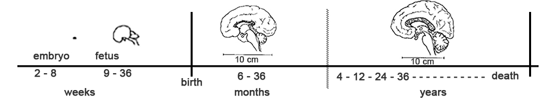
This timeline is just a guideline because all fetuses have slightly different neurodevelopment. Neurodevelopment also continues long after childbirth throughout a person’s life.
References and additional readings:
Budday, S., Steinmann, P., & Kuhl, E. (2015). Physical biology of human brain development. Frontiers in Cellular Neuroscience, 9. https://www.frontiersin.org/articles/10.3389/fncel.2015.00257
Fetal development: What happens during the 3rd trimester? (n.d.). Mayo Clinic. Retrieved May 9, 2023, from https://www.mayoclinic.org/healthy-lifestyle/pregnancy-week-by-week/in-depth/fetal-development/art-20045997
Stiles, J., & Jernigan, T. L. (2010). The basics of brain development. Neuropsychology Review, 20(4), 327–348. https://doi.org/10.1007/s11065-010-9148-4
Content collated and written by Jackson Pilifant, Reed’24.