In the previous post we described how the data is collected using Traction Force Microscopy (TFM). The process of imaging outputs several “movies,” which display the cells exerting forces and moving the beads embedded in the compliant gel matrix.
The data analysis algorithm is threefold: 1) tracking, 2) low-pass filtering, and 3) calculating traction stresses.
To calculate the displacements of the beads, a reference image of the beads on a plain compliant matrix is compared to an image of the beads on a compliant matrix with the cell on top of it which then pulls on the substrate near its edges. Using an array containing the tracked particle data for each frame of the movie, the displacements are extrapolated with stochastic drift taken into account. These data are then output onto an XY grid at each time interval.
Then, a low pass-filter is added to remove the high-frequency noise from the displacement data.
Next, the displacement data is correlated to the traction stresses through an algorithm derived by Style et. al. (2014) along with the elasticity theory, which states that the properties of the compliant matrix such as thickness, stiffness, and compressibility must be taken into account when considering traction stresses, which are continuous distributions of forces (Abidi, 2016). Modeling the gel as a “spring,” we can use Hooke’s Law, F=-kx, where k is the elasticity constant, x is displacement, and F is the force.
Finally, once the traction stresses have been computed, we will overlay a plot of the displacement and traction stress vectors on top of an image of the cell, as shown in Figure 1 (Abidi, 2016).
Figure 1: A force vector field calculated by Abidi (2016) using example data from Style et. al. (2014)

References
Abrar A. Abidi. Quantifying cellular mechanotransduction in morphogenesis and cancer. Reed College, 2016.
Robert W Style, Rotislav Boltyanskiy, Guy K German, Callen Hyland, Christopher W MacMinn, Aaron F Mertz, Larry A Wilen, Ye Xu, and Eric R Dufresne. Traction force microscopy in physics and biology. Soft Matter, 10(23):4047-4055, 2014.
One way we intend to examine the role of our target proteins, CG and Flapwing (flw) is by RNA interference knockdown (RNAi, in which the target proteins are inhibited in live cells, those cells exposed to the apical constriction signaling ligand, Fog, and the cells fixed so their response may be quantified. But in addition to knockdown, we also plan to measure target overexpression and localization. To these ends, we will use recombinant DNA techniques to produce clones of our target proteins’ coding sequences, in the form of a synthetic vector.
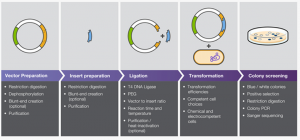
The process of producing a vector applicable to the proteins of interest first requires that template DNA of both targets are amplified by polymerase chain reaction (PCR). Once the DNA has been scaled up, it is precipitated and resuspended in water before being “digested” by two specialized restriction enzymes that sever the DNA at specific locations in sequences artificially inserted into the coding gene for the target proteins.

Because the cleavage sites on any two antiparallel sites cut by the same restriction enzyme will be complimentary, it is possible to place complimentary cut sites on both our insert and a cloning vector (pMT/V5-His A), thus allowing the cloning vector’s cut ends to match to the cut ends of our insert and for the two to bind. The process of combining vector and insert and encouraging them to bind and circularize is known as ligation.
Once ligation is complete and the insert DNA has been successfully added to the vector, the product is introduced to a bacterial culture and allowed to propagate before the culture is spun down by centrifuge and the DNA is isolated, for further use in increasing the expression of our target proteins in cell culture.

Due to difficulties with the cloning procedures, we are still currently attempting to successfully force the vector to take up the insert and vectorize the genes of our target proteins, which is necessary to introduce this increased protein expression into cell cultures. We have as such added CIP to the PMT His-A digestions, which prevents the vector from re-forming with itself and requires the binding of the insert for the DNA to circularize, and are expecting better results from our ligations in the future.
References:
PARF analysis or Permeabilization Activated Reduction in Fluorescence analysis allows researchers to describe the rate of dissociation of a fluorescently tagged protein of interest from intracellular structures. I will primarily be spending most of my time conducting PARF analysis to determine the effect of depleted SPECC1L on focal adhesions.
In our experiments, we transfect, or introduce, fluorescently tagged vinculin, which is one protein known to be found in focal adhesions. We visualize the focal adhesions using TIRF microscopy, and take control videos for each condition. Primarily, however, we are interested in the rate of dissociation of the fluorescently tagged vinculin from focal adhesions. This rate of dissociation corresponds to the size and relative strength of the focal adhesions. To cause this dissociation, we add digitonin to the cell media as we are filming to induce permeabilization of the cell membrane. To see more, look at the bottom of the post for a supplemental video!
This permeabilization results in an unbalanced gradient of fluorescent vinculin, with a high concentration within the cell, and a low concentration in the surrounding media. This gradient favors the dissociation of the vinculin into the external media. We measure the loss of fluorescence for around two minutes and then can fit this to an exponential decay. Using both a positive and negative control that causes larger and smaller focal adhesions, we can determine the effect of depleted SPECC1L on a cell’s focal adhesions.
Supplementary Material:
Supplemental Figure 1. This technique can be applied to various other proteins of interest. In the above video, we permeabilize these S2R+ cells expressing both fluorescent Naus GFP and fluorescent mCherry cortactin with digitonin after 20 seconds. We can then visualize the loss of fluorescent cortactin as it dissociates from the cell into the surrounding media.
Note: To view the video, you have to download it! If you have a Mac, click on the link while pressing Ctrl, and download the file.
Citations: