Team Force

Our team will use a physical approach to investigate the role of SPECC1L (Split Discs) in cellular contractility by quantifying force expression through Traction Force Microscopy (TFM).

In TFM, cells are adhered to compliant gel matrices with fluorescent beads. The traction forces are found as shown in Fig. 1

Figure 1: Measurement of cellular contraction in cells (Jacobs et al., 2012)

The movement of the fluorescent beads is tracked using the microscope, and along with the known physical characteristics of the substrate such as thickness and stiffness, we can extrapolate the force vectors between the cell and gel matrix.

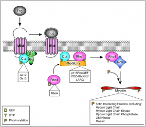
We are using gene inhibition using dsRNA induced gene silencing (RNAi) to target proteins such as Spaghetti Squash (Sqh), Myosin Binding Subunit (Mbs), and our protein of interest, SPECC1L. We will compare the force expression of Split Discs against that of Sqh depleted cells, which will cause the inactivation of non-muscle Myosin II (NMII) and hypocontractile cells, as well as Mbs depleted cells, which will result in the absence of dephosphorylation of RLC, causing an open NMII and hypercontractility (Abidi, 2016).

References:

Abrar A. Abidi. Quantifying cellular mechanotransduction in morphogenesis and cancer. Reed College, 2016.

Christopher R. Jacobs, Hayden Huang, and Ronald Y Kwon. Introduction to Cell Mechanics and Mechanobiology. Garland Science, 2012.

CG/Flapwing NMII

In order to form many of the essential structures of the body, such as blood vessels and the gut, a tissue must be able to fold upon itself into a tube. The process to create these structures is driven by cell-shape change, also known as morphogenesis. The folded gastrulation (Fog) pathway is one way cells can be induced to change shape. When a Fog ligand binds to its target receptor, the cytoskeletal network made up of actin and non-muscle myosin II induces constriction of the cell apices, causing the tissue to fold. Through cooperative investigation with computational biologists, a selection of fourteen target proteins with predicted involvement in the Fog pathway and its associated mechanisms were identified and tested for their significance in this pathway by their inhibition in cultured Drosophila melanogaster cells, using RNA-interference knockdown.

Figure 1. The Folded Gastrulation Pathway [Manning et al., 2014]. The Fog Pathway is initiated by the secreted ligand Fog binding to the heterotrimeric G-protein coupled receptor “Mist”, releasing previously-inactive Concertina (Cta) from the two trimer components G? and G? so it may bind to RhoGEF2. The binding of RhoGEF2 causes the small GTPase Rho1 to activate Rho kinase (Rok), which then phosphorylates the NMII regulatory light chain “Spaghetti Squash” (Sqh) and induces contraction of the cell’s apical actomyosin network (Manning et al., 2014).

Preliminary study of the fourteen candidate proteins showed that two in particular, CG and Flapwing (flw), exhibited significantly reduced contractility in knockdown and are therefore the focus of our study. While nothing is known or documented regarding the protein CG, flw has been thus far observed to act as a phosphorylating agent of Sqh and therefore an effector of NMII, similar to the known activities of Rho (Figure 1). Flw is also known to encode the β isoform of phosphatase protein type 1 (PP1), a protein that is highly conserved among all animals (Kirchner et al., 2007).

References:

  1. Manning A.J., Rogers S.L. 2014. The Fog signaling pathway: Insights into signaling in morphogenesis. Developmental Biology. 394(1): 6-14.
  2. Kirchner J., Gross S., Bennett D., Alphey L. 2007. The Nonmuscle Myosin Phosphatase PP1β (flapwing) Negatively Regulates Jun N-Terminal Kinase in Wing Imaginal Discs of Drosophila. Genetics. 175(4):1741-1749.


SPECC1L

Cellular migration is an essential feature of the development of tissues, organs, and overall facial morphogenesis. SPECC1L encodes a protein that functionally interacts with actin and microtubules, two key components of the cell cytoskeleton. Mutations in SPECC1L observed in humans, zebrafish, and Drosophila, reveal that SPECC1L plays an essential role in the development of a “face”, through the closure of the neural tube, and its role in cranial neural crest cell delamination. Cranial neural crest cell (CNCC) delamination describes the migration of CNCCs from the embryonic neural folds to the pharyngeal arches, thereby helping to form key features of the embryo face.

This delamination implies the role of SPECC1L in cellular contractility and migration. Non-muscle myosin II plays a critical role in the development of cellular protrusions, known as lamellipodia, necessary to the process of cell migrations. Focal adhesions form in the lamellipodia, and mature in the lamella, which is characterized by thicker bundles of actin and slower retrograde flow. These focal adhesions are critical to the ability of the cell to form attachments to the extracellular matrix (the substrate on which it crawls).

Figure 1. The above figure depicts the different types of attachments that the cytoskeleton and cell can make to the extracellular matrix. While disassembly can occur at any point within the maturation of the nascent adhesions in the lamella, the formation of focal adhesions and fibrillar adhesions is critical to pulling the cell forward.

The role of non-muscle myosin II in the formation of these cellular adhesions to the extracellular matrix suggests that it may be affected by the depletion of SPECC1L. Students in both biology and physics will collaborate on this research project to ultimately develop a greater understanding of the association between non-muscle myosin II and SPECC1L.

References:

  1. L. Gfrerer, V. Shubinets, T. Hoyos, Y. Kong, C. Nguyen, P. Pietschmann, C.C. Morton, R. L. Maas, E.C. Liao. Functional analysis of SPECC1L in craniofacial development and oblique facial cleft pathogenesis. Plast. Reconstr. Surg., 134 (2014), pp. 748-759
  2. Vicente-Manzanares, M., Ma, X., Adelstein, R. S. & Horwitz, A. R.Non-muscle myosin II takes centre stage in cell adhesion and migration. Nature Rev. Mol. Cell Biol. 10, 778– 790 (2009).
  3. Saadi I, Alkuraya FS, Gisselbrecht SS, Goessling W, Cavallesco R, Turbe-Doan A, et al. Deficiency of the cytoskeletal protein SPECC1L leads to oblique facial clefting. Am J Hum Genet (2011) 89(1):44–55. doi:10.1016/j.ajhg.2011.05.023
  4. Wilson, N. R., Olm-Shipman, A. J., Acevedo, D. S., Palaniyandi, K., Hall, E. G., Kosa, E., et al. (2016). SPECC1L deficiency results in increased adherens junction stability and reduced cranial neural crest cell delamination. Sci. Rep. 6:17735. doi: 10.1038/srep17735