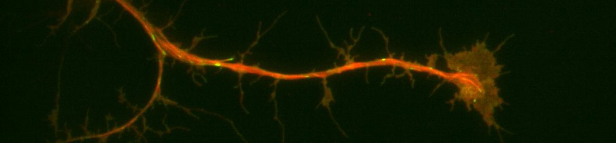
One aspect of my thesis is exploring co-localization of Split Discs with other proteins in drosophila cells. In order to do this, not only does wet lab work need to be accomplished, but mathematical analysis (in this case using Mander’s coefficient).
Fluorescence microscopy does not have the ability to see whether or not two molecules are directly interacting. However, by looking to see if they co-localize in the cell, it can be determined whether they interact with the same complexes in the cell. The limit for fluorescence microscopy is the resolution of the images produced. Because of this, small numbers of puncta are not sufficient for determining whether or not the experimental molecules are actually co-localized. Multiple puncta from different regions within the cell must be used in analysis so the data is not limited to overlapping puncta which are a result of organelles that are close in proximity to one another.
In order to quantitatively determine the correlation of co-localization in the cell, mathematical analysis of the data is employed. For my thesis, I am employing Mander’s Overlap Coefficient (MOC) for this analysis because it does not require distinguishing fluorescence as being the result of a fluorescent protein or background noise. MOC is able to do this because it only compares the co-occurrence of fluorescence among pixels. MOC = ∑i(Ri×Gi) / √(∑iR2i×∑iG2i) where Ri and Gi are the average level of grey from the red and green fluorescence respectively (Manders et al., 1993). MOC has a range of 0 – 1 and Ri and Gi have a range of -1 – +1. The limitation to this equation is that the ratio of values can result in ambiguous numbers. Therefore, the numerator and denominator can be split up in such a way to account for the ambiguity. From this we get two coefficients: M1 (fraction of red fluorescence in areas with green fluorescence) and M2 (fraction of green fluorescence in areas with red fluorescence) (Manders et al., 1993). M1 = (∑iRi,colocal) / ∑iRi where Ri,colocal = Ri if Gi > 0 and Ri,colocal = 0 if Gi = 0 and M2 = (∑iGi,colocal) / ∑iGi where Gi,colocal = Gi if Ri > 0 and Gi,colocal = 0 if Ri = 0 (Manders et al., 1993). The larger MOC, M1, and M2 are the stronger the evidence for co-localization of the proteins within a cell. In my thesis, MOC, M1, and M2, will be gathered for each cell to determine whether or not Split Discs are co-localizing with other specific proteins.
References:
Dunn, K. W., Kamocka, M. M., & McDonald, J. H. (2011). A practical guide to evaluating colocalization in biological microscopy. American Journal of Physiology – Cell Physiology, 300(4), C723–C742. http://doi.org/10.1152/ajpcell.00462.2010
Manders, E. M., Verbeek, F. J. & Aten, J. A. (1993). Measurement of co‐localization of objects in dual‐colour confocal images. Journal of Microscopy, 169, 375-382. doi:10.1111/j.1365-2818.1993.tb03313.x