BioID of SPECC1L

BirA is biotin protein ligase in E. coli that selectively adds biotin to a subunit of acetyl-CoA carboxylase. Roux et al. created a mutant BirA that isn’t selective to its native substrate and will instead biotinylate any proteins that are close-by. This allows for identification of protein-protein interactions in eukaryotic cells by creating a fusion protein of BirA and a protein of interest that will biotinylate proximal proteins, which can then be captured and identified. Protein identification utilizes the strong association of biotin and streptavidin (or avidin) to capture proteins that have been biotinylated by BirA, followed by mass spectroscopy to identify biotinylated proteins.


Figure 1. Schematic of BioID assay from Roux et al. 2012.

I am investigating the protein interactions of an actin-microtubule crosslinking protein coded by the gene SPECC1L (with an ortholog known as “split discs” in Drosophila). Mutations in SPECC1L have been identified in a number of diverse cases of orofacial cleft, and thus knowledge of protein-protein interactions by the product of SPECC1L is particularly important to understanding the developmental basis of this set of conditions.

To this end, I am using a BirA-split discs construct to investigate what proteins split discs interacts with in vivo in Drosophila. This construct will ideally biotinylate proteins that split discs typically interacts with, without excessive non-target biotinylation and without affecting the behavior or split discs. I will then capture biotinylated proteins using the biotin-streptavidin interaction and identify using mass spectrometry.


References:

Roux KJ, Kim DI, Raida M, Burke B. A promiscuous biotin ligase fusion protein identifies proximal and interacting proteins in mammalian cells. The Journal of Cell Biology. 2012;196(6):801.

CG and Flapwing: Knockouts, Knockdowns, and RNA Interference

When attempting to understand a protein’s function in a cell, the effects of removing that protein can be very telling. As such, knockdowns and knockouts, techniques which remove a targeted protein from acting in the cell, are widely used to identify how a cell behaves without the functions performed by said target protein.


A knockout is an irreversible procedure that removes the target protein from the cell permanently, often by editing the genome itself to either deactivate or directly remove the target protein’s coding sequence. This can be achieved by selective breeding when dealing with whole organisms, but when working with cell cultures, knockouts are typically accomplished by means such as the gene-editing kit TALEN or, in recent years, by use of the tool CRISPR-cas9.
A knockdown, on the other hand, is a repeated procedure which removes the target protein from the cell temporarily. By treating cells on a regular basis, the target protein is disrupted from its usual function, and the cells can be observed without its influence. If the treatment ceases, the target protein will no longer be disrupted and the cell will return to normal function, if mildly worse for wear. The distinction between these two procedures can be likened to ending a fight (knockout) as opposed to temporarily ‘gaining advantage’ or suspending the fight (knockdown).


In our experiment, we have performed a RNA interference (RNAi) knockdown of our target proteins flw and CG. This is achieved first by the transformation of genomic DNA (gDNA) that codes for the target protein into double stranded RNA (dsRNA), and then by simply exposing cultured cells to the dsRNA on a sustained and regular basis.
The reason this apparently simple method (which requires more pipetting and tube shuffling than that short sentence might imply) works and removes the target protein from action is due to the cell’s own inherent defensive mechanisms. When exposed to free floating dsRNA in solution, some dsRNA is naturally taken up into the cell, where it is recognized as foreign and chopped to pieces. Ironically, this causes the fragmented dsRNA to bind to messenger RNA (mRNA) already in the cell that matches its sequence, whereupon the entire dsRNA-fragment-mRNA amalgamation is recognized as foreign and chopped to pieces.


Figure 1. dsRNA suspended in cell media is taken into a cell, recognized as a foreign component, and cut into pieces by the appropriate enzymes. The cut pieces of dsRNA attach to pieces of mRNA naturally present in the cell, which are subsequently tagged for sheering due to their binding with a foreign component.


When the dsRNA is in sufficient concentration, this defensive response results in the cell being unable to translate the appropriate mRNA into the target protein, since it is instead destroying that mRNA as fast as it can. Once depleted of the target protein, cells can be treated, fixed, stained, or any combination thereof, and the effects of the knockdown can be observed to extrapolate the target protein’s function.

Team Force: Data Analysis Techniques

In the previous post we described how the data is collected using Traction Force Microscopy (TFM). The process of imaging outputs several “movies,” which display the cells exerting forces and moving the beads embedded in the compliant gel matrix.

The data analysis algorithm is threefold: 1) tracking, 2) low-pass filtering, and 3) calculating traction stresses.

To calculate the displacements of the beads, a reference image of the beads on a plain compliant matrix is compared to an image of the beads on a compliant matrix with the cell on top of it which then pulls on the substrate near its edges. Using an array containing the tracked particle data for each frame of the movie, the displacements are extrapolated with stochastic drift taken into account. These data are then output onto an XY grid at each time interval.

Then, a low pass-filter is added to remove the high-frequency noise from the displacement data.

Next, the displacement data is correlated to the traction stresses through an algorithm derived by Style et. al. (2014) along with the elasticity theory, which states that the properties of the compliant matrix such as thickness, stiffness, and compressibility must be taken into account when considering traction stresses, which are continuous distributions of forces (Abidi, 2016). Modeling the gel as a “spring,” we can use Hooke’s Law, F=-kx, where k is the elasticity constant, x is displacement, and F is the force.

Finally, once the traction stresses have been computed, we will overlay a plot of the displacement and traction stress vectors on top of an image of the cell, as shown in Figure 1 (Abidi, 2016).

Figure 1: A force vector field calculated by Abidi (2016) using example data from Style et. al. (2014)


References

Abrar A. Abidi. Quantifying cellular mechanotransduction in morphogenesis and cancer. Reed College, 2016.

Robert W Style, Rotislav Boltyanskiy, Guy K German, Callen Hyland, Christopher W MacMinn, Aaron F Mertz, Larry A Wilen, Ye Xu, and Eric R Dufresne. Traction force microscopy in physics and biology. Soft Matter, 10(23):4047-4055, 2014.


CG and Flapwing: creating clones using synthetic vectors

One way we intend to examine the role of our target proteins, CG and Flapwing (flw) is by RNA interference knockdown (RNAi, in which the target proteins are inhibited in live cells, those cells exposed to the apical constriction signaling ligand, Fog, and the cells fixed so their response may be quantified. But in addition to knockdown, we also plan to measure target overexpression and localization. To these ends, we will use recombinant DNA techniques to produce clones of our target proteins’ coding sequences, in the form of a synthetic vector.


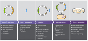
The process of producing a vector applicable to the proteins of interest first requires that template DNA of both targets are amplified by polymerase chain reaction (PCR). Once the DNA has been scaled up, it is precipitated and resuspended in water before being “digested” by two specialized restriction enzymes that sever the DNA at specific locations in sequences artificially inserted into the coding gene for the target proteins.

Because the cleavage sites on any two antiparallel sites cut by the same restriction enzyme will be complimentary, it is possible to place complimentary cut sites on both our insert and a cloning vector (pMT/V5-His A), thus allowing the cloning vector’s cut ends to match to the cut ends of our insert and for the two to bind. The process of combining vector and insert and encouraging them to bind and circularize is known as ligation.

Once ligation is complete and the insert DNA has been successfully added to the vector, the product is introduced to a bacterial culture and allowed to propagate before the culture is spun down by centrifuge and the DNA is isolated, for further use in increasing the expression of our target proteins in cell culture.

Due to difficulties with the cloning procedures, we are still currently attempting to successfully force the vector to take up the insert and vectorize the genes of our target proteins, which is necessary to introduce this increased protein expression into cell cultures. We have as such added CIP to the PMT His-A digestions, which prevents the vector from re-forming with itself and requires the binding of the insert for the DNA to circularize, and are expecting better results from our ligations in the future.


References:


  1. Molecular Cell Biology. 4th edition. Lodish H, Berk A, Zipursky SL, et al. New York: W.H. Freeman; 2000
  2. Thermo Fisher Scientific (2018). Traditional Cloning Basics | Thermo Fisher Scientific – US.

PARF Analysis

PARF analysis or Permeabilization Activated Reduction in Fluorescence analysis allows researchers to describe the rate of dissociation of a fluorescently tagged protein of interest from intracellular structures. I will primarily be spending most of my time conducting PARF analysis to determine the effect of depleted SPECC1L on focal adhesions.

In our experiments, we transfect, or introduce, fluorescently tagged vinculin, which is one protein known to be found in focal adhesions. We visualize the focal adhesions using TIRF microscopy, and take control videos for each condition. Primarily, however, we are interested in the rate of dissociation of the fluorescently tagged vinculin from focal adhesions. This rate of dissociation corresponds to the size and relative strength of the focal adhesions. To cause this dissociation, we add digitonin to the cell media as we are filming to induce permeabilization of the cell membrane. To see more, look at the bottom of the post for a supplemental video!

This permeabilization results in an unbalanced gradient of fluorescent vinculin, with a high concentration within the cell, and a low concentration in the surrounding media. This gradient favors the dissociation of the vinculin into the external media. We measure the loss of fluorescence for around two minutes and then can fit this to an exponential decay. Using both a positive and negative control that causes larger and smaller focal adhesions, we can determine the effect of depleted SPECC1L on a cell’s focal adhesions.

Supplementary Material:

PARF Blog Post Attachment

Supplemental Figure 1. This technique can be applied to various other proteins of interest. In the above video, we permeabilize these S2R+ cells expressing both fluorescent Naus GFP and fluorescent mCherry cortactin with digitonin after 20 seconds. We can then visualize the loss of fluorescent cortactin as it dissociates from the cell into the surrounding media.

Note: To view the video, you have to download it! If you have a Mac, click on the link while pressing Ctrl, and download the file.

Citations:

  1.  Singh PP, Hawthorne JL, Quintero OA. Permeabilization Activated Reduction in Fluorescence (PARF): a novel method to measure kinetics of protein interactions with intracellular structures. Cytoskeleton (Hoboken). 2016 June ; 73(6): 271–285. doi:10.1002/cm.21306.

Team Force

Our team will use a physical approach to investigate the role of SPECC1L (Split Discs) in cellular contractility by quantifying force expression through Traction Force Microscopy (TFM).

In TFM, cells are adhered to compliant gel matrices with fluorescent beads. The traction forces are found as shown in Fig. 1

Figure 1: Measurement of cellular contraction in cells (Jacobs et al., 2012)

The movement of the fluorescent beads is tracked using the microscope, and along with the known physical characteristics of the substrate such as thickness and stiffness, we can extrapolate the force vectors between the cell and gel matrix.

We are using gene inhibition using dsRNA induced gene silencing (RNAi) to target proteins such as Spaghetti Squash (Sqh), Myosin Binding Subunit (Mbs), and our protein of interest, SPECC1L. We will compare the force expression of Split Discs against that of Sqh depleted cells, which will cause the inactivation of non-muscle Myosin II (NMII) and hypocontractile cells, as well as Mbs depleted cells, which will result in the absence of dephosphorylation of RLC, causing an open NMII and hypercontractility (Abidi, 2016).

References:

Abrar A. Abidi. Quantifying cellular mechanotransduction in morphogenesis and cancer. Reed College, 2016.

Christopher R. Jacobs, Hayden Huang, and Ronald Y Kwon. Introduction to Cell Mechanics and Mechanobiology. Garland Science, 2012.

CG/Flapwing NMII

In order to form many of the essential structures of the body, such as blood vessels and the gut, a tissue must be able to fold upon itself into a tube. The process to create these structures is driven by cell-shape change, also known as morphogenesis. The folded gastrulation (Fog) pathway is one way cells can be induced to change shape. When a Fog ligand binds to its target receptor, the cytoskeletal network made up of actin and non-muscle myosin II induces constriction of the cell apices, causing the tissue to fold. Through cooperative investigation with computational biologists, a selection of fourteen target proteins with predicted involvement in the Fog pathway and its associated mechanisms were identified and tested for their significance in this pathway by their inhibition in cultured Drosophila melanogaster cells, using RNA-interference knockdown.

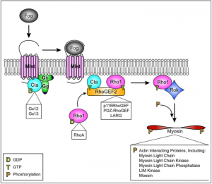
Figure 1. The Folded Gastrulation Pathway [Manning et al., 2014]. The Fog Pathway is initiated by the secreted ligand Fog binding to the heterotrimeric G-protein coupled receptor “Mist”, releasing previously-inactive Concertina (Cta) from the two trimer components G? and G? so it may bind to RhoGEF2. The binding of RhoGEF2 causes the small GTPase Rho1 to activate Rho kinase (Rok), which then phosphorylates the NMII regulatory light chain “Spaghetti Squash” (Sqh) and induces contraction of the cell’s apical actomyosin network (Manning et al., 2014).

Preliminary study of the fourteen candidate proteins showed that two in particular, CG and Flapwing (flw), exhibited significantly reduced contractility in knockdown and are therefore the focus of our study. While nothing is known or documented regarding the protein CG, flw has been thus far observed to act as a phosphorylating agent of Sqh and therefore an effector of NMII, similar to the known activities of Rho (Figure 1). Flw is also known to encode the β isoform of phosphatase protein type 1 (PP1), a protein that is highly conserved among all animals (Kirchner et al., 2007).

References:

  1. Manning A.J., Rogers S.L. 2014. The Fog signaling pathway: Insights into signaling in morphogenesis. Developmental Biology. 394(1): 6-14.
  2. Kirchner J., Gross S., Bennett D., Alphey L. 2007. The Nonmuscle Myosin Phosphatase PP1β (flapwing) Negatively Regulates Jun N-Terminal Kinase in Wing Imaginal Discs of Drosophila. Genetics. 175(4):1741-1749.


SPECC1L

Cellular migration is an essential feature of the development of tissues, organs, and overall facial morphogenesis. SPECC1L encodes a protein that functionally interacts with actin and microtubules, two key components of the cell cytoskeleton. Mutations in SPECC1L observed in humans, zebrafish, and Drosophila, reveal that SPECC1L plays an essential role in the development of a “face”, through the closure of the neural tube, and its role in cranial neural crest cell delamination. Cranial neural crest cell (CNCC) delamination describes the migration of CNCCs from the embryonic neural folds to the pharyngeal arches, thereby helping to form key features of the embryo face.

This delamination implies the role of SPECC1L in cellular contractility and migration. Non-muscle myosin II plays a critical role in the development of cellular protrusions, known as lamellipodia, necessary to the process of cell migrations. Focal adhesions form in the lamellipodia, and mature in the lamella, which is characterized by thicker bundles of actin and slower retrograde flow. These focal adhesions are critical to the ability of the cell to form attachments to the extracellular matrix (the substrate on which it crawls).

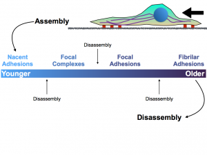
Figure 1. The above figure depicts the different types of attachments that the cytoskeleton and cell can make to the extracellular matrix. While disassembly can occur at any point within the maturation of the nascent adhesions in the lamella, the formation of focal adhesions and fibrillar adhesions is critical to pulling the cell forward.

The role of non-muscle myosin II in the formation of these cellular adhesions to the extracellular matrix suggests that it may be affected by the depletion of SPECC1L. Students in both biology and physics will collaborate on this research project to ultimately develop a greater understanding of the association between non-muscle myosin II and SPECC1L.

References:

  1. L. Gfrerer, V. Shubinets, T. Hoyos, Y. Kong, C. Nguyen, P. Pietschmann, C.C. Morton, R. L. Maas, E.C. Liao. Functional analysis of SPECC1L in craniofacial development and oblique facial cleft pathogenesis. Plast. Reconstr. Surg., 134 (2014), pp. 748-759
  2. Vicente-Manzanares, M., Ma, X., Adelstein, R. S. & Horwitz, A. R.Non-muscle myosin II takes centre stage in cell adhesion and migration. Nature Rev. Mol. Cell Biol. 10, 778– 790 (2009).
  3. Saadi I, Alkuraya FS, Gisselbrecht SS, Goessling W, Cavallesco R, Turbe-Doan A, et al. Deficiency of the cytoskeletal protein SPECC1L leads to oblique facial clefting. Am J Hum Genet (2011) 89(1):44–55. doi:10.1016/j.ajhg.2011.05.023
  4. Wilson, N. R., Olm-Shipman, A. J., Acevedo, D. S., Palaniyandi, K., Hall, E. G., Kosa, E., et al. (2016). SPECC1L deficiency results in increased adherens junction stability and reduced cranial neural crest cell delamination. Sci. Rep. 6:17735. doi: 10.1038/srep17735